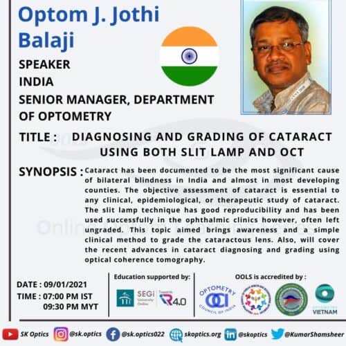
Mr. Jothi Balaji, a practicing optometrist, and researcher begin with a series of questions that help the audience to understand their level of knowledge in cataract grading. He introduces the need to learn about cataract grading by sharing that it has been the most common cause of preventable blindness globally for the past few decades. Mr. Balaji says that most individuals suffer through cataracts in their lives since it is an age-related condition.
He explains that most cataracts can be classified into three primary types depending on the area where the cataract starts to develop.
- Nuclear sclerosis – opacification occurs in the nucleus of the crystalline lens.
- Cortical spokes cataract – opacification occurs in the cortex capsule
- Posterior subcapsular cataract – opacification develops in the posterior capsule.
He explains the need to grade cataracts as the patients with functional vision loss associated with cataracts must undergo surgery.
We learn about the three commonly slit lamp illumination techniques used to grade cataracts.
- Diffuse illumination
- Optic section
- Retro-illumination
Mr. Jothi Balaji explains that when grading a cataract, LOCS 3 is the most commonly used method. Grading for different cataract types
- Nuclear sclerosis
- Cortical spokes cataract
- Posterior subcapsular cataract
We learn about the Posterior polar cataract in detail
Prevalence and risks
Clinical presentation
Clinical diagnosis
Classification and grading
Schroeder posterior polar cataract grading in children
Lastly, Mr. Balaji explains how anterior segment OCT is being used to diagnose and manage posterior polar cataracts. The session ends with an engaging question-and-answer session with the live audience.
References
Papers
Posterior Polar Cataract
Lee MW, Lee YC. Phacoemulsification of posterior polar cataracts–a surgical challenge. Br J Ophthalmol. 2003 Nov;87(11):1426-7.
https://pubmed.ncbi.nlm.nih.gov/14609852/
Vogt G, Puhó E, Czeizel AE. Population-based case-control study of isolated congenital cataract. Birth Defects Res A Clin Mol Teratol. 2005 Dec;73(12):997-1005.
https://pubmed.ncbi.nlm.nih.gov/16323161/
Kalantan H. Posterior polar cataract: A review. Saudi J Ophthalmol. 2012 Jan;26(1):41-9.
https://pubmed.ncbi.nlm.nih.gov/23960967/
Schroeder HW. The management of posterior polar cataract: the role of patching and grading. Strabismus. 2005 Dec;13(4):153-6.
https://pubmed.ncbi.nlm.nih.gov/16361185/
Chan TC, Li EY, Yau JC. Application of anterior segment optical coherence tomography to identify eyes with posterior polar cataract at high risk for posterior capsule rupture. J Cataract Refract Surg. 2014 Dec;40(12):2076-81.
