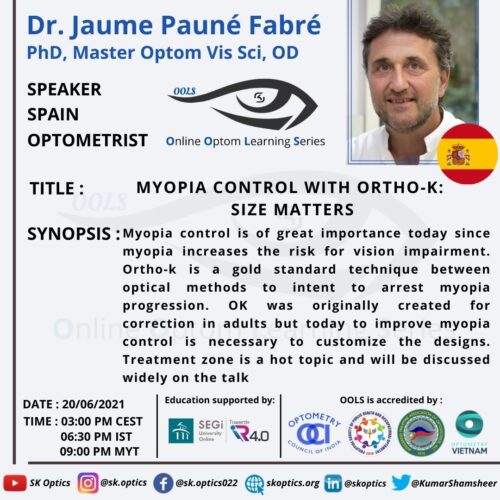
Dr. Jaume Pauné Fabré, a practicing optometrist, educator, and researcher, explains the need for myopia management. We briefly learn about myopia and orthokeratology.
- Myopia prevalence
- Myopia associated pathologies
- Orthokeratology and myopia management
- Orthokeratology optics
After understanding myopia control and orthokeratology basics, we learn about the different factors that can affect myopia control.
- Age and myopia control
- Pupil Size
- Higehr Order Aberration
- Imposed Defocus: Dose-dependent response
- Plus power ring
- Plus power ring and pupil size
- Obtaining a smaller treatment zone diameter
- Customizing lenses
- Monitoring Myopia
- When to start and stop myopia control
Dr. Jaume Pauné Fabré summarizes the talk with a few take-home points. The session ends with a question and answers segment for the live audience.
References
Papers
Dolgin E. The myopia boom. Nature. 2015 Mar 19;519(7543):276-8.
https://pubmed.ncbi.nlm.nih.gov/25788077/
Saw SM, Gazzard G, Shih-Yen EC, Chua WH. Myopia and associated pathological complications. Ophthalmic Physiol Opt. 2005 Sep;25(5):381-91.
https://pubmed.ncbi.nlm.nih.gov/16101943/
Cho P, Cheung SW, Edwards M. The longitudinal orthokeratology research in children (LORIC) in Hong Kong: a pilot study on refractive changes and myopic control. Curr Eye Res. 2005 Jan;30(1):71-80.
https://pubmed.ncbi.nlm.nih.gov/15875367/
Sun Y, Xu F, Zhang T, Liu M, Wang D, Chen Y, Liu Q. Orthokeratology to control myopia progression: a meta-analysis. PLoS One. 2015 Apr 9;10(4):e0124535.
https://pubmed.ncbi.nlm.nih.gov/25855979/
Wen D, Huang J, Chen H, Bao F, Savini G, Calossi A, Chen H, Li X, Wang Q. Efficacy and Acceptability of Orthokeratology for Slowing Myopic Progression in Children: A Systematic Review and Meta-Analysis. J Ophthalmol. 2015;2015:360806.
https://pubmed.ncbi.nlm.nih.gov/26221539/
Smith EL 3rd. Optical treatment strategies to slow myopia progression: effects of the visual extent of the optical treatment zone. Exp Eye Res. 2013 Sep;114:77-88.
https://pubmed.ncbi.nlm.nih.gov/23290590/
Queirós A, Lopes-Ferreira D, Yeoh B, Issacs S, Amorim-De-Sousa A, Villa-Collar C, González-Méijome J. Refractive, biometric and corneal topographic parameter changes during 12 months of orthokeratology. Clin Exp Optom. 2020 Jul;103(4):454-462.
https://pubmed.ncbi.nlm.nih.gov/31694069/
Chen Z, Niu L, Xue F, Qu X, Zhou Z, Zhou X, Chu R. Impact of pupil diameter on axial growth in orthokeratology. Optom Vis Sci. 2012 Nov;89(11):1636-40.
https://pubmed.ncbi.nlm.nih.gov/23026791/
Hiraoka T, Kotsuka J, Kakita T, Okamoto F, Oshika T. Relationship between higher-order wavefront aberrations and natural progression of myopia in schoolchildren. Sci Rep. 2017 Aug 11;7(1):7876.
https://pubmed.ncbi.nlm.nih.gov/28801659/
Benavente-Pérez A, Nour A, Troilo D. Axial eye growth and refractive error development can be modified by exposing the peripheral retina to relative myopic or hyperopic defocus. Invest Ophthalmol Vis Sci. 2014 Sep 4;55(10):6765-73.
https://pubmed.ncbi.nlm.nih.gov/25190657/
Wang J, Yang D, Bi H, Du B, Lin W, Gu T, Zhang B, Wei R. A New Method to Analyze the Relative Corneal Refractive Power and Its Association to Myopic Progression Control With Orthokeratology. Transl Vis Sci Technol. 2018 Nov 30;7(6):17.
https://pubmed.ncbi.nlm.nih.gov/30533280/
Pauné J, Fonts S, Rodríguez L, Queirós A. The Role of Back Optic Zone Diameter in Myopia Control with Orthokeratology Lenses. J Clin Med. 2021 Jan 18;10(2):336.
https://pubmed.ncbi.nlm.nih.gov/33477514/
Marcotte-Collard R, Simard P, Michaud L. Analysis of Two Orthokeratology Lens Designs and Comparison of Their Optical Effects on the Cornea. Eye Contact Lens. 2018 Sep;44(5):322-329.
https://pubmed.ncbi.nlm.nih.gov/29489498/
Jones LA, Mitchell GL, Mutti DO, Hayes JR, Moeschberger ML, Zadnik K. Comparison of ocular component growth curves among refractive error groups in children. Invest Ophthalmol Vis Sci. 2005 Jul;46(7):2317-27.
https://pubmed.ncbi.nlm.nih.gov/15980217/
González Blanco F, Sanz Ferńandez JC, Muńoz Sanz MA. Axial length, corneal radius, and age of myopia onset. Optom Vis Sci. 2008 Feb;85(2):89-96.
https://pubmed.ncbi.nlm.nih.gov/18296925/
He X, Zou H, Lu L, Zhao R, Zhao H, Li Q, Zhu J. Axial length/corneal radius ratio: association with refractive state and role on myopia detection combined with visual acuity in Chinese schoolchildren. PLoS One. 2015 Feb 18;10(2):e0111766.
https://pubmed.ncbi.nlm.nih.gov/25693186/
Tideman JWL, Polling JR, Vingerling JR, Jaddoe VWV, Williams C, Guggenheim JA, Klaver CCW. Axial length growth and the risk of developing myopia in European children. Acta Ophthalmol. 2018 May;96(3):301-309.
https://pubmed.ncbi.nlm.nih.gov/29265742/
Taneri S, Arba-Mosquera S, Rost A, Kießler S, Dick HB. Repeatability and reproducibility of manifest refraction. J Cataract Refract Surg. 2020 Dec;46(12):1659-1666.
https://pubmed.ncbi.nlm.nih.gov/33259390/
Rauscher FG, Lange H, Yahiaoui-Doktor M, Tegetmeyer H, Sterker I, Hinz A, Wahl S, Wiedemann P, Ohlendorf A, Blendowske R. Agreement and Repeatability of Noncycloplegic and Cycloplegic Wavefront-based Autorefraction in Children. Optom Vis Sci. 2019 Nov;96(11):879-889.
https://pubmed.ncbi.nlm.nih.gov/31703049/
Cho P, Cheung SW. Discontinuation of orthokeratology on eyeball elongation (DOEE). Cont Lens Anterior Eye. 2017 Apr;40(2):82-87.
https://pubmed.ncbi.nlm.nih.gov/28038841/
McBrien NA, Adams DW. A longitudinal investigation of adult-onset and adult-progression of myopia in an occupational group. Refractive and biometric findings. Invest Ophthalmol Vis Sci. 1997 Feb;38(2):321-33.
https://pubmed.ncbi.nlm.nih.gov/9040464/
Bullimore MA, Jones LA, Moeschberger ML, Zadnik K, Payor RE. A retrospective study of myopia progression in adult contact lens wearers. Invest Ophthalmol Vis Sci. 2002 Jul;43(7):2110-3.
