January 16, 2021
| No Comments

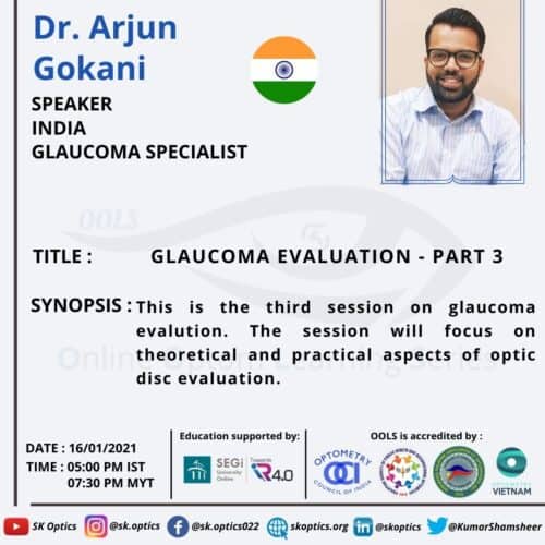
Dr. Arjun Gokani, an ophthalmologist and a glaucoma expert, begins by stating that optic disc evaluation is one of the three main pillars of glaucoma diagnosis. He starts by explaining with a video on to perform a 90D/78D on a slit-lamp. He then talks about all the elements that need to be considered when evaluating the optic disc.
The five R’s of optic disc evaluation
- Scleral ring – the outermost ring of the optic disc
- Neuro-retinal rim – retinal nerve fiber arrangement
- ISNT rule
- Vertical cup/disc ratio
- Disc damage likelihood scale
- Retinal nerve fiber layer – the entire top layer of the retina
- Retinal or optic disc hemorrhages – mainly seen in patients with normal-tension glaucoma
- Region of peri-papillary atrophy – the atrophy around the optic disc
Dr. Gokani ends the session by discussing some specific clinical scenarios, which includes
- Bayonetting sign
- Laminar dotsign/shadow sign
- Focal atrophy and polar notching
- Advanced glaucomatous cupping/ bean pot cupping
- Asymmetry of cups
The session ends with an interesting discussion with the host.
More Relatable Blogs:
- https://visionscienceacademy.org/evaluation-of-island-of-vision-relics-of-a-bygone-age/
- https://visionscienceacademy.org/optometrists-and-glaucoma/
- https://visionscienceacademy.org/24-2c-new-modality-of-visual-field-analyzer/
- https://visionscienceacademy.org/novel-techniques-towards-visual-field-testing-in-paediatric-patients/
- https://visionscienceacademy.org/change-of-ocular-dimensions-in-different-types-of-glaucoma/
