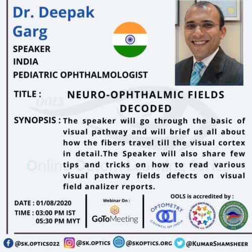
Dr. Deepak Garg begins with a brief introduction to the optic nerve pathway. He starts from the retinal fibers to the visual cortex. He talks in detail about the retinal fiber positioning along the visual pathway and the importance of understanding these arrangements for an accurate diagnosis. He graphically illustrates the nerve fiber arrangements at the following locations:
1. Papillomacular Bundle
2. Optic Disc
3. Optic Nerve
4. Optic Chiasm & Willbrand’s Knee
5. Optic Tract 6. Lateral Geniculate Body
7. Optic Radiations
8. Visual Cortex
Dr. Deepak also explains the anatomy of the occipital cortex and the blood supply at the occipital cortex and then describes the terminology used to explain field defects. This is followed by an in-depth analysis of the different field defects and how various field defects can help us determine where is the neuro-ophthalmic defect.
He gives some tips about how different field defects follow specific rules, which can be used effectively to determine the defect location.
Dr. Garg also explains how to differentiate between neuro-ophthalmic field defects and other field defects.
Lastly, Dr. Garg gives some essential clinical tips while answering the audience questions during the question-and-answer session.
