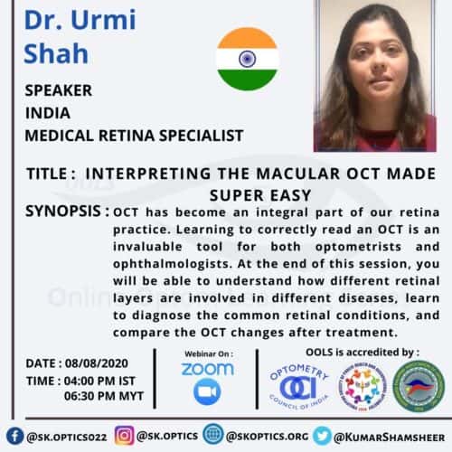
Dr. Urmi Shah starts with the basics of optical coherence tomography (OCT) and the importance of OCT interpretation for all eye care practitioners. OCT is similar to ultrasound, except it uses light waves. Dr. Shah specifies, in short, the working of the OCT machine. Then, she introduces the different OCT technologies available.
She talks briefly about OCT acquisition procedures, the types of scans, advantages, and disadvantages. We learn that on an OCT report, the three retinal nuclear layers appear hyporeflective (dark), and the other layers appear hyperreflective (white).
We learn about the different locations where changes in the retina may occur and also the site of fluid collection. Changes in OCT are visualized through changes in reflectivity. Only fluid collection can cause hyporeflectivity, causes of hyperreflectivity changes are numerous. Then Dr. Shah walks us through the OCT interpretation for sign and symptoms of common retinal conditions, which includes
- Age-related macular degeneration (AMD)
- Dry AMD
- Drusen
- Geographic atrophy
- Wet AMD
- Choroidal neovascular membrane
- Pigment epithelial detachment
- Disciform scaring
- Dry AMD
- Vitreoretinal Interface Disorders
- Vitreomacular adhesion
- Vitreomacular traction
- Macular hole
- Epiretinal membrane
- Cystoid macular edema
- Diabetic retinopathy
- Venous Occlusions
- Pseudophakic cystoid macular edema
- Central serous retinopathy
- Arterial Occlusions
- Tractional retinal detachment
- Solar retinopathy
Dr. Shah also shows images of extramacular lesions captured on OCT. She not only shows OCT images for all these conditions, but she also adds it’s clinical uses. She enlists steps to interpret the OCT systematically.
We hear about different OCT machines, programs, and other clinical insights to enhance the use of OCT for patient care. Lastly, she talks about the recent most advances of OCT angiography and intraoperative OCT. The session is concluded with a question and answer session with the audience.
